About the Author
Katrina Furth, Ph.D. Associate Scholar, Charlotte Lozier Institute and SAGI Senior Advisor
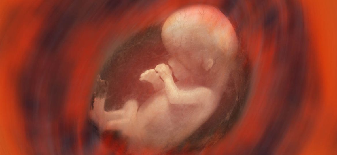
Seeking God’s Fingerprints in the Development of Human Life
Words of Wisdom: “For you created my inmost being; you knit me together in my mother’s womb. I praise you because I am fearfully and wonderfully made” (Psalm 139:13-14 NIV).
Life is sacred at every stage. From the moment the sperm fuses with a mother’s egg and creates a single-celled unique human individual, to the earliest stirrings of consciousness in the womb, to the pinnacle of decision-making in adulthood, the journey of neurological development is a testament to the inherent dignity and value of every human life.
WHY IS HUMAN LIFE SACRED?
There are two clear reasons. First, God set human beings apart from the rest of creation to reflect and bear His image. When God created human beings in His own image, He bestowed in us a little mark of the divine that makes us irreplaceable and worthy of dignity and respect. Each person is God’s image-bearer from the moment of conception. There is enough genetic variation that no two humans have been, or ever will be, genetically identical (1). Even in the case of identical twins, there are small genetic differences arising from some sections of DNA getting copied more than once, called copy number variations (2). Secondly, human life is made sacred by the fact that God loves every individual discretely. Jesus explained this in the parable of the lost sheep in Matthew 18:12-14: “If a man has a hundred sheep and one of them wanders away, what will he do? Won’t he leave the ninety-nine others on the hills and go out to search for the one that is lost? And if he finds it, I tell you the truth, he will rejoice over it more than over the ninety-nine that didn’t wander away! In the same way, it is not my heavenly Father’s will that even one of these little ones should perish.”
God gives each human life value as He winds together our DNA and knits collagen, blood, and bone together in the womb. God loves every child, the child born into welcoming arms and the child aborted in a quiet hospital room.
MYSTERIES OF HUMAN DEVELOPMENT
As we start growing from a single cell, layers and structures form and fold like a glorious origami. How do the cells all become different types of tissue, assemble into the correct structures, and work together to serve our body and sustain life? Why does the cornea of the eye develop transparently, while the whites of our eyes block incoming light? Why does each person have a differently shaped outer ear, but everyone has a single ear canal that connects to the eardrum and inner ear? How do the genetic instructions we’re given not make mistakes, such as creating extra inner ear bones or mispositioning those bones so that sounds are not amplified, even in the womb? The mysteries multiply, especially when dealing with a complex system such as the human brain. The infant brain has about 100 billion neurons, each forming thousands of connections with other neurons. We cannot come close computationally to modeling the brain with its myriad and ever-changing connections, let alone the development of each and every neuron! The mystery of fetal development points like a spotlight to God the Creator.
DEVELOPING A BRAIN THAT SENSES
Our abilities to perceive unfold within the sanctity of the womb. The ability to sense our body in space, hear, smell, see, and feel originates in the miraculous complexity of the developing brain. Understanding the mysterious journey of neurological development proves valuable.
The brain starts forming as the neural tube in the fifth gestational week. From this simple start, the top regions of the neural tube expand and intricately fold. Brain regions and structures develop in place with divine precision, guided by chemical messages. By the midpoint of gestation, around 5 ½ months, the brain assumes its adult shape. The next few months see its once smooth surface adorned with intricate contours, increasing its surface area to accommodate more neurons (3).
Neural stem cells give rise to a vast array of neurons and glial cells, each destined to fulfill a unique role in the symphony of thought and sensation. From deep inside the brain, nascent neurons migrate, sculpting the cortex, where their birthdate dictates their destiny. Chemical messages pass between neurons, directing the connections that underpin cognition (4). Remarkably, signs of electrical activity have been recorded as early as 8 weeks’ gestation (5). As pregnancy progresses, more neural circuits rapidly form, laying the groundwork for future cognitive abilities.
But the journey does not end with birth. Rather, it is a lifelong odyssey of growth and refinement. Like a sculptor refining a masterpiece, the brain undergoes a process of blooming and pruning, shedding redundant connections to sculpt a more efficient neural architecture. For example, a fetus at 28 weeks gestation has more neurons than he will ever have again, as unused neurons get removed (6). Similarly, two-year-olds have more synapses than most adults. However, these extra synapses appear redundant and are pruned during development. Part of the reason that toddlers teeter is that multiple neurons try to control a single muscle, and these neurons do not coordinate their efforts (7).
Fetal experiences play an important role in sensory development. For example, sensations that a baby can practice in the womb, like taste, hearing and touch are better developed than senses like seeing. A baby may hear his mother’s voice and taste foods from her diet in the amniotic fluid, but very little light penetrates the uterus (8). This contributes to the fact that babies are born legally blind (9), but they can recognize their mother’s native languages (10).
Fetal experiences play an important role in sensory development. For example, sensations that a baby can practice in the womb, like taste, hearing and touch are better developed than senses like seeing. A baby may hear his mother’s voice and taste foods from her diet in the amniotic fluid, but very little light penetrates the uterus (8). This contributes to the fact that babies are born legally blind (9), but they can recognize their mother’s native languages (10).
DEVELOPING A SENSE OF PAIN
Furthermore, the preborn baby can feel pain by 15 weeks’ gestation and possibly even earlier (11). As touch receptors develop, starting near the mouth and hands at 7 ½ weeks gestation, they immediately start forming connections with the nascent spinal cord (12). In fact, the embryo will reflexively move away from a touch at 7 ½ weeks (13). Neurotransmitters dedicated to pain, such as substance P and enkephalin appear at 10 to 12 weeks and 12 to 14 weeks respectively (14). Leading neuroscientists conclude that neural structures for processing pain are functional as early as 12 weeks’ gestation (15). Babies respond to painful procedures with a stress response by 18 weeks gestation (16), and can be observed crying out in the womb during an anesthetic injection as early as 23 weeks gestation (17). Finally, the younger a premature baby is, the greater their brain response to a painful heel lance, suggesting that both the fetus and premature children are more sensitive to pain than full-term newborns and adults (18). Taken together, a number of compelling studies demonstrate that the developing fetus is sensitive to pain inside the womb.
DEVELOPING A SENSE OF TASTE
Additionally, the preborn child starts tasting early in pregnancy. Nascent taste buds start forming on the tongue around 8 weeks’ gestation, about a month after a mother might first know she is pregnant. For these taste buds to work, they must connect to a specialized sensory nerve to relay the taste information to the brain. Through an intricate interplay of chemical messages between cells, a taste bud only matures after it has appropriately connected to one of these nerves (19). The fetus can taste around 4 months after conception (20). Now, you’ve probably never considered what amniotic fluid might taste like, but you spent 5 months of your life drinking nothing but amniotic fluid! When a mother eats a meal – any meal – the flavors from the meal arrive in the amniotic fluid about 45 minutes later (21). The preborn child swallows more amniotic fluid if it is sweet and less if it is bitter (22). The flavors that a fetus tastes in the womb will even influence her food preferences when she later starts solid foods. For example, babies whose mothers drank carrot juice regularly during their 8th month of pregnancy made happier facial expressions while eating carrot-flavored cereal when they started eating solid foods (23).
When I was pregnant with my first daughter, my husband and I went to a restaurant with a reputation for really spicy food. Eating that meal was like wrestling with jalapenos. But I had never considered the effect that it would have on my daughter. About 20 minutes into the meal, she started thrashing inside of me, and kicking my bladder with a desperation and determination that I had not yet experienced! She basically performed over an hour of flips, kicks, and fetal karate moves until I decided that I would never again eat spicy food while she was inside me! To this day, my daughter refuses to eat anything spicy.
DEVELOPING A BRAIN THAT DECIDES
Every twitch, every impulse, and every decision originate in the miraculous complexity of the developing brain. At 14 weeks’ gestation, groundbreaking research revealed a remarkable discovery: the fetus exhibits purposeful movements, directing gestures more slowly toward her own eyes, mouth, and even the uterine wall (24). Prior to this milestone, fetal movements were characterized by jerky motions, lacking intentionality. However, at this stage, hands exhibit a newfound precision, slowing down as they approach their target. Intriguingly, if the fetus shares the womb with a twin, some movements are directed towards the sibling, executed with surprising gentleness (25). By 18 weeks, this dexterity becomes even more pronounced, with the fetus displaying quicker and more precise gestures towards her own features, particularly when utilizing her dominant hand (26).
THE BEATING HEART
Every living human has a beating heart by six weeks gestation. Specifically, by 22 days after fertilization, the heart starts to pump blood rhythmically (27). The embryonic heart beats faster than the mother’s heart from the sixth week onwards. While an average adult heart rate is about 60 to 100 beats per minute, the embryo’s heart beats at 110 beats per minute at 6 weeks gestation and 170 bpm at 10 weeks gestation (28). This means that by the end of the 6th gestational week, the heart will have already beat over 1 million times, and by birth this same heart will beat 54 million times (29).
The preborn child needs a heart so that it can grow. In fact, the heart is the first organ to function in the developing human embryo (30). Vitally, it circulates nutrients and oxygen-rich blood once the embryo can no longer meet its nutritional needs by diffusion from the placenta alone (31).
At six weeks’ gestation, the heart starts as two tubes, which fuse together and fold during the sixth week, forming an s-curve. Even at these early stages, the heart begins to form primitive heart valves to prevent the backflow of blood and help push the blood forward through the rest of the body (32). In the seventh week, the two upper sections become the atria, and in the eighth week the larger, stronger ventricles form. The heart reaches its final shape by the tenth week, with two atria, two ventricles, and circulatory blood vessels, although these blood vessels mostly bypass the liver and lungs to help the embryo get oxygenated blood from the umbilical cord to the rest of the body (33).
BETTER THAN BELIEF
My friend Noor was an independent business woman with her own dance studio. She loved to dress up and stay out late dancing! Her positive pregnancy test left her feeling upset and confused. Babies are inconvenient. They don’t let you go out all night. As her son’s heart started beating, Noor fought off waves of nausea. As her son’s started making intentional movements, Noor became more limited in dance instruction. Some people advised Noor to get an abortion; but Noor chose life. Her son was born two days before my daughter in the same hospital. Noor babysat for me all the time, and the two kids are good friends. I can’t imagine a world without her son’s laughter. But Noor’s very hip lifestyle was permanently altered by raising a child. Her chic apartment became littered with kid toys, wet burp cloths, and unmatched socks. Noor’s courageous sacrifices make her one of the most pro-life people I've ever met.
Being pro-life is about acting as the hands and feet of Jesus to love the least, the lost, and the lonely. Spending time in awe and wonder at the precision of neurological development in the womb is very important, as it guides us to stand in awe of our creator, and recognize that life is sacred at every stage; but that is not enough. God has called us all to love the woman who gets to nurture a sacred life in her womb, and support both lives with dignity and value.
REFERENCES AND NOTES
- National Institutes of Health (US) and Biological Sciences Curriculum Study, Understanding Human Genetic Variation, NIH Curriculum Supplement Series. (National Institutes of Health (US), 2007), https://www.ncbi.nlm.nih.gov/books/NBK20363/.
- Carl E.G. Bruder et al., “Phenotypically Concordant and Discordant Monozygotic Twins Display Different DNA Copy-Number-Variation Profiles,” American Journal of Human Genetics 82, no. 3 (March 3, 2008): 763–71, https://doi.org/10.1016/j.ajhg.2007.12.011.
- Konkel Lindsey, “The Brain before Birth: Using FMRI to Explore the Secrets of Fetal Neurodevelopment,” Environmental Health Perspectives 126, no. 11 (2018): 112001, https://doi.org/10.1289/EHP2268.
- Kristin Keunen, Serena J. Counsell, and Manon J. N. L. Benders, “The Emergence of Functional Architecture during Early Brain Development,” NeuroImage, Functional Architecture of the Brain, 160 (October 15, 2017): 2–14, https://doi.org/10.1016/j.neuroimage.2017.01.047.
- Borkowski, Winslow J., and Richard L. Bernstine. “Electroencephalography of the Fetus.” Neurology 5, no. 5 (May 1, 1955): 362. https://doi.org/10.1212/WNL.5.5.362.
- Rachel Marsh, Andrew J. Gerber, and Bradley S. Peterson, “Neuroimaging Studies of Normal Brain Development and Their Relevance for Understanding Childhood Neuropsychiatric Disorders,” Journal of the American Academy of Child & Adolescent Psychiatry 47, no. 11 (November 1, 2008): 1233–51, https://doi.org/10.1097/CHI.0b013e318185e703.
- https://lozierinstitute.org/dive-deeper/brain-connections-for-movement-and-sensations/
- https://lozierinstitute.org/dive-deeper/the-newborn-senses-taste-smell/
- https://lozierinstitute.org/dive-deeper/the-newborn-senses-sight-and-eye-color/
- https://lozierinstitute.org/dive-deeper/the-newborn-senses-hearing/
- https://lozierinstitute.org/dive-deeper/prenatal-stress-and-pain/
- Humphrey, Tryphena. “Some Correlations between the Appearance of Human Fetal Reflexes and the Development of the Nervous System.” In Progress in Brain Research, edited by Dominick P. Purpura and J. P. Schadé, 4:93–135. Growth and Maturation of the Brain. Elsevier, 1964. https://doi.org/10.1016/S0079-6123(08)61273-X.
- Ibid.
- Bellieni, Carlo V. “Analgesia for Fetal Pain during Prenatal Surgery: 10 Years of Progress.” Pediatric Research, September 24, 2020, 1–7. https://doi.org/10.1038/s41390-020-01170-2.
- Derbyshire, Stuart WG, and John C Bockmann. “Reconsidering Fetal Pain.” Journal of Medical Ethics 46, no. 1 (January 2020): 3–6. https://doi.org/10.1136/medethics-2019-105701.
- Giannakoulopoulos, X., Teixeira, J., Fisk, N., & Glover, V. (1999). Human fetal and maternal noradrenaline responses to invasive procedures. Pediatric research, 45(4), 494-499.
- Bernardes, L. S., Rosa, A. S., Carvalho, M. A., Ottolia, J., Rubloski, J. M., Castro, D., … & de Andrade, D. C. (2021). Acute pain facial expressions in 23-week fetus. Ultrasound in Obstetrics & Gynecology. https://doi.org/10.1002/uog.23709 and https://lozierinstitute.org/wpcontent/uploads/2022/08/pr9_2020_11_25_diandrade_painreports-d-20-0059_sdc1b.mp4
- Marco Bartocci et al., “Pain Activates Cortical Areas in the Preterm Newborn Brain,” PAIN 122, no. 1 (May 1, 2006): 109–17, https://doi.org/10.1016/j.pain.2006.01.015.
- Witt, M., and K. Reutter. “Embryonic and Early Fetal Development of Human Taste Buds: A Transmission Electron Microscopical Study.” The Anatomical Record 246, no. 4 (December 1996): 507–23. https://doi.org/10.1002/(SICI)1097-0185(199612)246:4<507::AID-AR10>3.0.CO;2-S.
- https://lozierinstitute.org/dive-deeper/fetal-taste-and-maternal-diet/
- Mennella, J., Johnson, A. and Beauchamp, G. “Garlic Ingestion by Pregnant Women Alters the Odor of Amniotic Fluid,” Chemical Senses 20, no. 2 (April 1, 1995): 207–9, https://doi.org/10.1093/chemse/20.2.207.
- Liley, A. W. “The Foetus as a Personality.” The Australian and New Zealand Journal of Psychiatry 6, no. 2 (June 1972): 99–105. https://doi.org/10.3109/00048677209159688.
- Mennella, Julie A., Coren P. Jagnow, and Gary K. Beauchamp. “Prenatal and Postnatal Flavor Learning by Human Infants.” Pediatrics 107, no. 6 (June 2001): E88. https://doi.org/10.1542/peds.107.6.e88.
- Umberto Castiello et al., “Wired to Be Social: The Ontogeny of Human Interaction,” PLOS ONE 5, no. 10 (October 7, 2010): e13199, https://doi.org/10.1371/journal.pone.0013199.
- Ibid and https://lozierinstitute.org/fetal-development/week-14/
- Valentina Parma et al., “The Origin of Human Handedness and Its Role in Pre-Birth Motor Control,” Scientific Reports 7, no. 1 (December 1, 2017): 16804, https://doi.org/10.1038/s41598017-16827-y.
- Asp, Michaela, Stefania Giacomello, Ludvig Larsson, Chenglin Wu, Daniel Fürth, Xiaoyan Qian, Eva Wärdell et al. ‘A spatiotemporal organ-wide gene expression and cell atlas of the developing human heart.’ Cell 179, no. 7 (2019): 1647-1660. https://doi.org/10.1016/j.cell.2019.11.025
- Papaioannou, G. I., Syngelaki, A., Poon, L. C., Ross, J. A., & Nicolaides, K. H. (2010). Normal ranges of embryonic length, embryonic heart rate, gestational sac diameter and yolk sac diameter at 6–10 weeks. Fetal diagnosis and therapy, 28(4), 207-219. https://doi.org/10.1159/000319589.
- EHD: Appendix. ‘Appendix | Prenatal Overview.’ Accessed April 3, 2020. https://www.ehd.org/dev_article_appendix.php
- Tan C, M, J, Lewandowski A, J: The Transitional Heart: From Early Embryonic and Fetal Development to Neonatal Life. Fetal Diagn Ther 2020;47:373-386
- Hill, M.A. (2023, January 28),Cardiovascular System Development, Available at: https://embryology.med.unsw.edu.au/embryology/index.php/Cardiovascular_System_Develop ment
- Combs MD, Yutzey KE. Heart valve development: regulatory networks in development and disease. Circ Res. 2009 Aug 28;105(5):408-21. doi: 10.1161/CIRCRESAHA.109.201566. PMID: 19713546; PMCID: PMC2777683.
- Buijtendijk, MFJ, Barnett, P, van den Hoff, MJB. Development of the human heart. Am J Med Genet Part C. 2020; 184C: 7– 22. https://doi.org/10.1002/ajmg.c.31778.
What an inspiring article! It made me look back on my pregnancies and relive all those beautiful live creating moments that God has blessed me with twice! This is an amazing article to share with those that do not understand that being pro life is not about telling a women what to do with her body but understanding the science behind how a fetus develops and how quickly life is formed. It may change the minds of those who are pro choice!